
|
Frullania rostrata (Hook. f. & Taylor) Gottsche, Lindenb. & Nees
 |
|
|
Family: Frullaniaceae
[Frullania diplota Taylor, moreFrullania megalocarpa Taylor, Frullania minutistipula Steph., Frullania novaustralis S.Hatt., Jungermannia rostrata Hook.f. et Taylor, Neohattoria australis R.M.Schust., Schusterella australis (R.M.Schust.) S.Hatt., Sharp et Mizut.]  John E. Braggins |
Brief description: Frullania rostrata is a polymorphic species, especially in the size of the plant, form of the lobuli, underleaves and styli, as well as cell anatomy. For example, shoot size ranges from less than 400 µm wide to more than 1mm wide, form of the underleaf varies from those with entire lateral margins to those with small teeth or angulations, and lobe apices vary from rounded to appendiculate. In fact, with respect to herbarium material, some forms of F. rostrata with acute to appendiculate leaf apices have been mistaken for F. apiculata; von Konrat & Braggins (2001) provided a key to help distinguish between these two species. See: von Konrat, M. and Braggins, J. E. (2001). Notes on Five Frullania Species from Australia, including Typification, Synonyms, and New Localities. The Journal of the Hattori Botanical Laboratory. 91: 229-263. Affinities, differentiation & variation: This species is most similar to Frullania pauciexiliens, but plants of the latter are typically smaller, monoicous, have a 3-keeled perianth with an ornamented surface, have fewer elaters per capsule (only 12 to 22), and the first branch leaf (BL1) is frequently reduced in size and atypical compared to normal stem leaves, with all three segments explanate or at most sulcate, and leaves characteristic of the main stem starting at either BL2, BL3 or BL4. In contrast, F. rostrata is typically dioicous, has a 3-keeled perianth without any surface ornamentation, and has a greater number of elaters (24 to 42). The BL1 is usually similar to those of normal stem leaves i.e. 1 explanate dorsal lobe + 1 saccate lobule + 1 stylus. Close morphological affinities of F. rostrata alsolie with the F. junghuhniana species-complex and they share the following assemblage of characteristics: 1) underleaves typically entire; 2) lobules usually large (its area obscuring 1/3–1/2 the exposed area of the dorsal lobe), 1.5–2× long as wide, and obliquely spreading; 3) styli generally large in comparison to the lobule; 4) distinctly 3-keeled perianth; 5) often reddish-brown pigmentation; and 6) extremely variable in form, including some very small phenotypes. Frullania rostrata can immediately be separated from one variant of the F. junghuhniana species-complex, F. junghuhniana var. minutissima, because F. rostrata lacks themammillose lobules which are a distinctive feature of this variety. In most cases, a diagnostic character associated with the anatomy of the lobule can be used to separate F. rostrata from members of the F. junghuhniana species-complex. In both species, at approximately one-third from the lobule mouth there is a ± discoloured, gibbous, protuberant cell: In F. rostrata this cell is usually equal in length to surrounding cells or at most only slightly longer. In members of the F. junghuhniana species-complex this cell is usually conspicuously longer (to 3.25×) than adjacent cells and up to 30µm in length. Also, at an ultra structural level the surface of the inner capsule wall easily distinguishes between the two species: In F. rostrata the cell surface is densely covered with fine perforations and in F. junghuhniana the cell surface has 1–6 large distinct pores per cell, forming deep indentations, to 3.0 µm long × 2.5 µm wide. The confusion between these two species is highlighted by Hattori (1972b) who described F. junghuhniana subsp. wattsii as a new subspecies, but later in 1979 reduced it to synonymy under F. rostrata. Frullania rostrata is a polymorphic species, especially in the size of the plant, form of the lobuli, underleaves and styli, as well as cell anatomy. For example, shoot size ranges from less than 400 µm wide to more than 1mm wide, form of the underleaf varies from those with entire lateral margins to those with small teeth or angulations, and lobe apices vary from rounded to appendiculate. In fact, with respect to herbarium material, some forms of F. rostrata with acute to appendiculate leaf apices have been mistaken for F. apiculata; von Konrat & Braggins (2001) provided a key to help distinguish between these two species [see under F. (subg. Frullania) apiculata]. The size of the stylus also varies enormously in F. rostrata and can easily cause confusion if not well developed. Some phenotypes, often found in Tasmania, have an exceptionally large stylus, at least 50 cells in total, and almost equal in length to the lobule; in contrast, styli of this stature are rarely encountered in typical forms of the species elsewhere. Also of note, is a form of F. rostrata, here recognised as only a coastal phenotype, with almost squarrose lobes when wet and dry, slightly larger oil-bodies and slightly larger median cells.
In New Zealand, very small forms of F. rostrata may be mistaken for F. truncata, but the large rectangular stylus with a truncate apex, immediately distinguishes this taxon from F. rostrata. Furthermore, F. rostrata never has a conspicuous protruding cell that develops just above the mouth of the lobule, which appears to be a feature of species of sect. Amphijubula. Chemically, the two species are very different from each other with sesquiterpene lactones present in F. rostrata and absent in F. truncata. General distribution: Found throughout Australasia including, New Zealand where it occurs on all the major islands, i.e., North Island, South Island, and Stewart Island; Australia and Tasmania. Detailed description: Plants small to medium, main shoots (380) 480–900 (1050) µm wide, forming olive-green, copper-brown, to black patches, closely to loosely adhering to substrate. Leading stem 10–30 (60) mm long and 60–150µm in diam., 6–9 cells wide, little differentiation between cortical cells (16–25 in no.) and medullary cells (18–30 in no.), occasionally former slightly smaller than the latter, both with firm walls, lumen irregularly shaped. Branching often regularly pinnate, occasionally 2 or 3-pinnate, branches with progressively smaller leaves. Dimorphic branching, with both Frullania-type (FB) and occasionally Lejeunea-type (LB) branching. Initial appendages of FB: First branch underleaf (BUL1) always with three distinct segments, the ventral lamina divided for ⅓–⅔ its length into two unequally or subequally sized lobes + 1 dorsal saccate lobe; First branch leaf (BL1)usually± characteristic of normal stem leaves (i.e.BL1: 1 explanate dorsal lobe + 1 saccate lobule + 1 stylus), only rarely are the appendages of BL1 reduced in size and elobulate with leaves characteristic of the main stem starting at BL2–3. Initial appendages of LB: BUL1 and BL1–3 are always reduced in size and lobule-free, formation of normal lobulate leaves occurring thereafter. Stemleavesof main branchflat when dry and wet, slightly imbricate to contiguous, suborbicular to broadly oval, (160) 180–580 (650) µm long × (90) 120–350 (400) µm wide with incurved distal margins, and dorsal margins extending beyond the farther edge of the stem, rounded apices and non-auriculate at the base, margins entire and surface smooth. Lobules ± remote from the stem (lobule attached to stem by 2–3(4) cells) and at angles of (20) 30–50 (60)° with the stem so that lobules tilted outwards; lobules similar in colour to other organs; cylindrically pitcher-shaped (orbicular in cross-section with up to 28 cells) and 1.5–2× long as wide; large (its area obscuring at 1/3–1/2 the exposed area of the dorsal lobe), (110) 130–210 (240) µm long × (60) 70–110 (130) µm wide; somewhat dorsiventrally compressed near mouth as compared to the gibbous upper half, the opening wide, extending along the abaxial lobule margin; ca. 1/3 from lobule mouth there is usually a ± discoloured, gibbous, protuberant cell, to 25 µm long and the wall up to 5µm thick; free margin of lobular mouth crenulate-sinuate, usually hyaline near mouth, lobule apex obtuse. Stylus medium to large, (5) 10–35 (55) cells in total, to 175 µm long × 125 µm wide, subtriangular to foliaceous with a rounded, subacute or acute apex, occasionally with a slime papilla at apex. Underleaves of leading stems1/3 to ½ the size of leaf lobes, distant, contiguous, or just overlapping with lobules, underleaves contiguous to distant from each other, usually almost as long as wide, occasionally slightly wider than long, (150) 180–330 (400) µm long × (100) 140–300 (400) µm wide, (1) 2–3.5 (5)× the stem in width, broadest at middle (8–40 cells wide); lateral margins usually entire, some rare phenotypes with small angulations or with 1 pair of very small teeth on the lateral margins; apex of underleaf bilobed to 1/2 its length, lobes 4–18 cells wide at base, rounded, acute to subacute apices, and separated by a U to V-shaped sinus. Rhizoid-initial area present near base of underleaf, rhizoids often seen, subhyaline, in short bundles. Plants either not microphyllous at all or exhibiting a pseudo-microphyllous habit with lobules of secondary stems ± similar size to main stem, but lobes and underleaves of secondary branches markedly smaller than those of leading stems. Lobe marginal cells ± rectangular, hyaline walls subequally thickened, cell cavities brownish red, to 8 µm long × 7 µm wide; median cells ± subquadrate to rectangular, hyaline walls subequally thickened, intermediate thickening rare to absent, wall thickness to 2.75 µm wide (without intermediate thickenings), cell cavities of median cells brownish red, (9) 11–15 (18) µm long × (5) 7–10 (11) µm wide, (1) 1.2–1.8 (2.5)× long as wide; cells becoming gradually larger basally, cavities of the basal median group of cells (16) 19–25 (30) µm long × (7) 10–14 (19) µm wide; walls of basal cells with subnodulose trigones and intermediate thickenings, walls and cavities brownish red. Median cells of underleaves ± subequally thickened so that the hyaline trigones and intermediate thickenings become indistinct, cell cavities to 14 µm long × 10 µm wide. Median cells oflobuleslightly longer than wide, cell cavities to 12 µm long × 7 µm wide, walls flexuose with indistinct trigones and small nodulose intermediate thickenings from the base of the lobule to the apex (both often heavily pigmented, olive-brown to dark brown in contrast to hyaline walls of the lobe and stem underleaf). Oil-bodies of the leaf lobe median cells: (2) 3–4 (5) oil-bodies per cell, typically small, spherical (1) 2–3 (4) µm in diam. to ovoid or ellipsoidal (2) 3–5 (6) µm × (1) 2–3 (4) µm, subhyaline, without any significant, visible, internal structure i.e. giving the appearance of being almost homogeneous. The oil-bodies are often similar in size or slightly smaller than the chloroplasts; occasionally the oilbodies are larger than the chloroplasts. The oil-bodies of the lobule and underleaf are similar to those encountered in the leaf lobe. Oil-bodies becoming progressively larger towards the basal cells: usually (2) 3–5 (6) per cell, ocelli never present, (3) 4–7 (10) µm long × (2) 3–5 (6) µm wide, coarsely granular to finely botryoidal. Asexual reproduction occasionally present via caducous leaf lobes and less often lobules, which develop propagula before they fall, or rarely via ± discoid gemmae from the leaf lobe margins. Plants usually dioicous (some phenotypes monoicous). Androecia subspherical to discoid, to 500 µm long × 450 µm wide, 2–4 (6) pairs of bracts, sessile (stalk with no leaf lobes) or terminal on very short-stalked branches (stalk with 1–4 vegetative leaf lobes). Gynoecia terminal on main or leading stem. A shoot system usually arising immediately below the set of 3–4 progressively larger bracts below the gynoecium or perianth (subfloral branch), occasionally arising one or more vegetative leaf-cycles below the gynoecia or perianth bracts i.e. a subfloral branch, or rarely occurring between bracts and perianth (subfloral innovation). ♀ bracts and bracteoles occasionally in pairs of 2 cycles but typically in pairs of 3–4 cycles, which distally, gradually increase in size and complexity. Innermost bract unequally bilobed; bract-lobe larger, ovate to oblong, narrowed toward acute apex, the margin irregularly entire, bract-lobule smaller, ovate–lanceolate, subacute, occasionally with 1 stylar tooth at the base; innermost bracteole free from bracts, oblong in outline with subparallel margins, about 1/2 bilobed, the sinus narrowed, lobes acute at apex, entire or with 1–2 short but distinct teeth at the base. Marginal cells of bract and bracteole ± subequally thickened, but towards the median cells, trigones becoming large and bulging. One or two archegonia per gynoecium. Perianth freely emergent, to 1200 µm long × 600 µm wide, exterior smoothly trigonous, oblong-ovate, tapering towards the apex into a short beak; perianth beak cylindrical, to 80 µm long, with a smooth mouth but the inner beak surface densely covered with large single-celled protuberances (to 10 µm long). Median cells of perianth with small triangular trigones and intermediate thickening rare or absent. Seta freely emergent, to 160 µm at widest axis; valves of capsule to 560 µm long × 280 µm wide; elaters (24) 26–34 (42) in total (arrangement on two alternating valves: 1+3+4 or 2+4+2), unispiral, 320µm long × 16 µm wide, elater surface irregularly rugose-granulate. Epidermal cell layer (11) 13–17 (19) cells at the widest region, the corner thickenings of the epidermal cells extend toward the centre of the tangential face for a short distance, as short rounded lobes and the juxtaposed corner thickenings form 3–4 lobed figures. Inner cell layer withthe thickenings appearing as a network-like configuration across the face; surface with ridges along the individual cells. Surface ultrastructure of the inner cell layer with fine granular perforations. Spores globose, 40–50 µm at widest axis, which is interspersed with 6–9 rosettes; each rosette 3–5 µm in diam., and bearing a ring of 7–9 ± conspicuous primary projections; the primary projections 0.75–2.0 µm high × 0.5–1.5 µm wide at base (often with a 1.5–2:1 length to width ratio), and taper gradually to a rounded or subacute apex, without divisions of any kind and almost never bearing secondary deposits; the spore wall papillae otherwise densely distributed between rosettes. Notes: No original material collected from the AucklandIslands around the time of 1844 to the Antarctic expedition exists in NY, G, BM, only material from later collections and publications, e.g. Flora of New Zealand and material collected by Colenso. Material from hb S, was selected as the lectotype because the label data matched the original protologue ‘Lord Auckland group; on Parmelia enteromorpha.’ Stephani (1892) stated that F. minutissima was a mixture of both F. falciloba and F. rostrata. However, examination of the lectotype from WELT (selected above), which was contained within a larger packet labeled “F. sp. .nov. 1886, # Col. 1381; F. minutissima,” within this packet are actually three “micro” packets – all containing F. rostrata. In one of these smaller packets a specimen of F. rostrata contained a perianth, which Colenso referred to in his original description and hence this entity was selected as the lectotype of F. minutissima. The larger packet also contained F. incumbens and F. aterrima. Hattori (1978b) reduced Frullania congesta (Hook. & Tayl.) Hook. & Tayl. to a synonym of F. rostrata. In this study, we have examined the type material and live specimens of F. congesta, and conclude that F. congesta should in fact be recognised as a distinct species. Therefore, we have removed it from synonymy with F. rostrata; formal reinstatement of F. congesta, coupled with a detailed description, will be published in a forthcoming revision of the New Zealand Frullania taxa. The key below provides a categorical distinction between the two species. Note, only a fragment of material is required with a stem and a single branch. ------------------------------------------------------------------------- 1.First branch leaf (BL1)usually± similar to those of the of normal stem leaves (i.e. BL1: 1 explanate dorsal lobe + 1 saccate lobule + stylus)............................................................................................... F. rostrata 2.First branch leaf (BL1) atypical of normal stem leaves (i.e. BL1: 1 explanate dorsal lobe + 2 saccate lobules + stylus......................................................................................................................... F. congesta ------------------------------------------------------------------------------- Despite the combination of morphological characters that usually separate F. rostrata from F. junghuhniana, there appears to be an interface, in subtropical Australia, where for some specimens it is difficult to precisely distinguish with certainty between the two species. It is in this region that extensive collections are required, and examination of a suite of morphological characters, including oil-bodies , and biochemical and molecular data is necessary to ascertain the taxonomic relationship between these two species.
It is also noteworthy that at least in New Zealand, forms of F. rostrata with angulations or small teeth on the lateral margins of the underleaf, have been found in co-existence with F. aterrima and these may be an indication of possible hybridisation between these two species. Hattori, S. 1979a. A revision of the Australasian species of the Frullania, Hepaticae, I. Journal of the Hattori Botanical Laboratory 45: 323-363. Hattori, S. 1979b. A revision of the Australasian species of the genus Frullania, Hepaticae, II. Journal of the Hattori Botanical Laboratory 46: 119-153. Hattori, S. 1983. A revision of the Australasian species of the genus Frullania, Hepaticae, III. Journal of the Hattori Botanical Laboratory 54: 133-182. Schuster, R.M. 1970. Studies on Antipodal Hepaticae, III. Jubulopsis Schuster, Neohattoria Kamimura and Amphijubula Schuster. Journal of the Hattori Botanical Laboratory 33: 266-304. von Konrat, M. and Braggins, J. E. (2001). Notes on Five Frullania Species from Australia, including Typification, Synonyms, and New Localities. The Journal of the Hattori Botanical Laboratory. 91: 229-263. von Konrat, M., Braggins J., Asakawa Y., & Masao (2006). Recognition of Frullania congesta: A case study to present a species concept and a synthesis of significant taxonomic characters for the large liverwort genus Frullania (Frullaniaceae). Journal of the Hattori Botanical Laboratory. 100: 553-576. |
 Matt von Konrat  Matt von Konrat  Matt von Konrat  Matt von Konrat  Matt von Konrat  Matt von Konrat  Matt von Konrat                        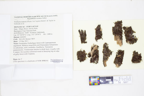 ")               ")                       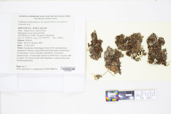          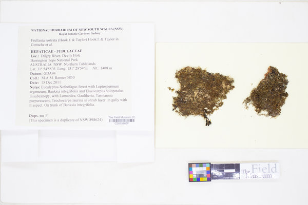    ")    ")            |
|

![]()
![]()
![]()
![]()