
|
Frullania knightbridgei von Konrat & de Lange
 |
|
|
Family: Frullaniaceae
 Matt von Konrat |
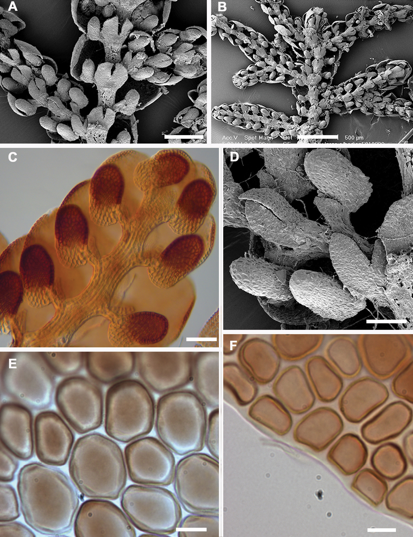
Description: Plants small to medium (main shoots to 600 µm wide), forming olive-green, copper-brown, to black patches, closely to loosely adhering to substrate. Leading stem 15–25 mm long and to 90µm in diameter, 6–9 cells wide, little differentiation between cortical cells (18–24 in no.) and medullary cells (14–28 in no.), the former often slightly smaller than the latter, both with firm walls, lumen irregularly shaped. Branching often regularly pinnate, occasionally bipinnate to rarely tripinnate, branches with progressively smaller leaves; Frullania-type branching. First branch underleaf (BUL1) always with three distinct segments, the ventral lamina divided for 1/3–2/3 its length into two unequally or subequally sized lobes + 1 dorsal saccate lobe; First branch leaf (BL1)usually± characteristic of normal stem leaves (i.e. 1 explanate dorsal lobe + 1 saccate lobule + 1 stylus). Stemleavesof main branchflat when dry and wet, slightly imbricate to contiguous, suborbicular to broadly ovate, to 375 µm long × 350 µm wide with incurved distal margins, dorsal margins extending beyond the farther edge of the stem, rounded apices, non-auriculate and ± subtruncate at the base, entire margins, smooth dorsal surface. Lobules remote from the stem (lobule attached to stem by 3–4 cells) and usually almost parallel with the stem so that the long axis of the lobule is ± parallel with the main stem (or at most lobules at angles of up to ca. 25° with the stem so that lobules only very slightly tilted outwards); lobules often bicoloured with the basal 2–5 cells towards the mouth (up to 0.25 of the lobule) hyaline to subhyaline, in contrast to the olive-green to brown pigmentation elsewhere; cylindrically helmet-shaped (orbicular in cross-section with up to 25 cells in circumference); lobules ± medium (lobule area obscuring no more than 0.25 × the exposed area of the dorsal lobe), ca. 1.75–2 × long as wide, 110–200 µm long × 60–100 µm wide (up to 12–14 cells high × 6–8 cells wide); ± equally inflated throughout (so that the sides of the lobule are ± parallel), the opening wide, extending only slightly along the abaxial lobule margin; ca. 2/3 from lobule apex there is usually a ± discoloured, gibbous, cell (the free margin of the cell with a heavily thickened wall); mouth nearest the stylus, truncate at base then cells with septa between adjacent cells ± swollen, the mouth thus then becoming crenulate-sinuate; lobule usually hyaline near mouth, lobule apex obtuse, surface of lobule smooth. Stylus medium in size (1/3–2/3× the length of the lobule), ± triangular, up to 60 µm long × 50 µm wide, (4) 5–6 (7) cells wide at base, (10) 12–24 (30) cells in total, occasionally with a poorly developed slime papilla at the apex.Underleaves of leading stems bilobed, obovate to rotundate, at most only contiguous with lobules, underleaves contiguous to distant from each other, usually long as wide, occasionally slightly longer than wide, (2) 2.5–3.5 (4)× the stem in width, to 100–175 µm long × 100–150 µm wide, broadest at middle, free lateral margins always entire; apex of underleaf bilobed to 1/3–½ its length, lobes separated by a V-shaped sinus, the lobes 9–14 cells wide at base and with blunt to subacute or rounded apices. Rhizoid-initial area present near base of underleaf, rhizoids often seen, subhyaline, in bundles, to 400 µm long. Not strictly microphyllous, lobules of secondary stems ± similar size to main stem, but lobes and underleaves of secondary branches slightly smaller than those of leading stems. Leaf-lobe: to 20 cells long, from base to apex, by 35 cells at widest region; with a band of conspicuously enlarged cells originating from the lobe base and extending out towards the lobe apex 10–12 cells, and up to 6 cells wide at the widest region. Lobe marginal cells ± rectangular to subquadrate, small to 8 µm long × 6 µm wide, hyaline walls subequally thickened, cell cavities brownish red. Cells of the middle region of the lobe are ± dimorphic in size; Type One [see below]: 4–6 rows of median cells, cells to 30 µm long × 22.5 µm wide (usually 2–2.75 × long as wide), thus similar in size to basal cells; Type Two [see below]: cells gradually becoming reduced in size (median cells to 15 µm long × 10 µm wide, usually 1.25–2 × long as wide, between central band of enlarged cells and lobe margin). Both cell types usually pentagonal or hexagonal, hyaline walls subequally thickened, intermediate thickening rare to absent, wall thickness to 2.75 µm wide, cell cavities of median cells brownish red. Cells becoming gradually larger basally, cavities of the basal cells to 40 µm long × 25 µm wide; walls of basal cells with small indistinct trigones and semi-straight walls without any intermediate thickenings, walls and cavities brownish red. Median cells of underleaves vary in shape and size, cells with heavily equally-thickened walls so that the hyaline trigones and intermediate thickenings become indistinct. Median cells oflobule as long as wide or slightly longer than wide, cell cavities to 14 µm long × 9 µm wide; cells near lobule mouth, irregular in shape with flexuose walls, indistinct trigones and occasional small nodulose intermediate thickenings; towards the lobule apex, cells gradually becoming more regular in shape, quadrate to rectangular and the cell walls becoming semi-straight. Oil bodies of lobe median cells dimorphic. Type One: (1)2(3) per cell, very large, occasionally spherical (2) 3–7 (9) µm in diam. but usually ovoid or ellipsoidal (5) 6–11 (13) × (4) 5–10 (12) µm, finely granular, occurs from basal cells through to central region of the lobe, occupying 3/4 to almost the entire cell lumen. Type One oil bodies larger than chloroplasts (if chloroplasts present at all). Type Two: 2–3 (4) oil bodies per cell, typically small, spherical (1) 2–4 (5) µm in diam. to ovoid or ellipsoidal (2) 3–5 (6) µm × (1) 2–3 (4) µm, subhyaline, without any significant, visible, internal structure i.e. giving the appearance of being almost homogeneous; these oil bodies often similar in size or slightly smaller than chloroplasts, occasionally slightly larger than chloroplasts. Oil bodies of lobule and underleaf of Type One. Asexual reproductionnot recognized. Plants dioecious, male plants slightly smaller than female plants. Androecia subspherical to spicate, 2–4 (6) pairs of bracts, terminal, usually on very short-stalked branches arising from the main stem, or occasionally from secondary branches (stalk with (1) 2–3 (4) vegetative leaf lobes). Gynoecia terminal on main or leading stem often bearing a subfloral innovation arising 3–4 bract-pair cycles back from the perianth or gynoecia. Innermost bract unequally bilobed; bract-lobe, lobule and innermost bracteole all with entire margins. Bract-lobe ovate to oblong, narrowed toward the rounded or subacute-acute apex, bract-lobule ovate–lanceolate, subacute; innermost bracteole free from bracts, oblong-ovate to oblong-obovate, ca. 1/2 bilobed, lobes convex-sided, subacute at apex, entire margins. Marginal cells of bract and bracteole ± subequally thickened, but towards the median cells, trigones becoming large and bulging. One archegonium per gynoecium. Perianth freely emergent, 900 µm long × 500 µm wide, perianth plicate, 1–2 dorsal keels + 2 lateral keels + 1–2 ventral keels, smooth surface, oblong-ovate, tapering towards the apex into a short beak; perianth beak cylindrical, with a smooth mouth but the inner beak surface densely covered with large single-celled protuberances. Walls of epidermal layer of capsule wall nodular where the lobes extend irregularly for a short distance over the tangential face toward the centre of the cell and have intermediate thickenings near the middle of the longer walls. Spores globose, 35–45 µm at widest axis, spore wall papillae densely distributed, equatorial face interspersed with 8–10 rosettes, 2.5–3 µm wide in the equatorial diameter, bearing a ring of 7–10 conspicuous primary projections, 0.75–1.5 µm long × 0.5–1.0 µm wide at base (often with a 1.5–2:1 length to width ratio), tapering gradually to a rounded apex, never papillate or bearing secondary short branches. This species is currently known from only four collections; two from the shore margin of Paterson Inlet, Stewart Island and two from the Auckland Islands. Frullania knightbridgei probably has a more extensive distribution than is currently known and it is quite likely that it resides unrecognized in New Zealand herbaria filed as a form of Frullania rostrata. Nevertheless, it would appear that Frullania knightbridgei is a species of southern distribution and it would be interesting to see if the distribution extends south to the Campbell Islands of the New Zealand botanical region; further field work is required to establish if the species grows on the other two main islands of the New Zealand archipelago, North and South Islands, it should for example be looked for along the Fiordland and Foveaux Strait coastline of the southern South Island. Note the type of Frullania pseudomeyeniana is a high elevation taxon at 1, 100 m whereas the New Zealand taxon is seemingly restricted to or near the shoreline or low elevation. Frullania knightbridgei is noteworthy in comparison with the majority of Frullania species in New Zealand for it appears to be a salt tolerant species. This is clearly evident in Stewart Island/Rakiura where Frullania knightbridgei was growing on twigs of Dracophyllum immediately adjacent the shoreline, at a height of about 50 cm above the sea. It was evident that for at least some periods of the year, this represented a harsh coastal environment where significant exposure to salt spray from violent wave action would be common.Elsewhere, Frullania ericoides reportedly develops in rock crevices exposed to sea in the Madeiran archipelago (Sim-Sim and Sergio 1992), and Schuster (1992) reported a variety of Frullania kunzei growing in mature mangrove swamp forest where salt spray and even rare submersion was possible. The high rainfall of the region, providing fresh water, is possibly a critical factor as Engel and Schuster (1973) described for tidal zone liverworts in southern Chile. Interestingly however, Engel and Schuster (1973) noted for Stewart Island a notable “lack of any Hepaticae in the intertidal zone where sea spray is a factor”. At least in the New Zealand botanical region, it is clear this is a habitat area that has traditionally been underexplored for liverworts. It would also be interesting to perform glasshouse experiments to investigate test the extent of salt tolerance in these organisms. Many critical morphological features have often been neglected in liverwort systematics (Schuster 1992; von Konrat et al. 1999, 2001a, 2006b), and scores of studies have been restricted to herbarium material where ephemeral structures; e.g., sporophytes and oil bodies, have not been available (von Konrat et al. 2006a, b; Heinrichs et al. 2009). The new species is morphologically aligned to a group of species representing Frullania subg. Microfrullania, which has been resolved as a monophyletic group (Hentschel et al. 2009). Inclusion of Frullania knightbridgei in Frullania subg. Microfrullania is also supported by molecular evidence as discussed above. Frullania subg. Microfrullania represents a clade with the most historical confusion out of all Frullania subgenera with taxa occurring in southern South America, Australasia and islands of the South Pacific, New Guinea, and Indonesia (von Konrat et al. 2006a, 2010). The new species appears almost to lie intermediate between Frullania rostrata, of New Zealand and Australia, and Frullania pseudomeyeniana S. Hatt. of New Caledonia. The latter is only known from the type material (New Caledonia, Mont Mou, N of Paita, 1100 m., Kitagawa 21422, NICH), which was examined by the senior author. Frullania knightbridgei also has some similarity with Frullania magellanica (Spreng.) F. Weber et Neesof Chile. Frullania knightbridgei superficially strongly resembles some forms of Frullania rostrata in plant size, the large styli and lobules, and the entire underleaves. However, with fresh material, Frullania knightbridgei is immediately discernable from Frullania rostrata by the presence of large oil bodies (usually only 2 per cell, occasionally 1 or 3) that almost occupy the entire cell lumen of basal and median cells of the leaf lobe. In the absence of oil body data, careful consideration has to be given to the anatomy of the leaf-lobe and -lobule to help differentiate between these species. In Frullania knightbridgei, cells towards the lobule apex progressively develop a more regular shape (quadrate to rectangular) and the cell walls become semi-straight. Conversely, the cell walls of both Frullania rostrata and Frullania pseudomeyeniana areflexuose with indistinct trigones, and with small, nodulose intermediate thickenings throughout the lobule, from the base to the apex. The unique cell anatomy of the leaf lobe in Frullania knightbridgei furtherplaces it into an isolated position within subg. Microfrullania; this species is seemingly unique in having a group of conspicuously enlarged cells, originating from the base of the lobe and extending 10–14 cells out toward the apex, forming a partial band or pseudo vitta up to 4–6 cells wide. The cells are enlarged to accommodate the typically 2 large oil bodies. The features of the oil bodies are unique within Frullania subg. Microfrullania. In those species examined thus far, the oil bodies of the leaf lobe median cells number from 2–4(5) per cell, are of small size and lack any significant ornamentation, almost appearing as homogeneous oil droplets (von Konrat et al. 2006a, 2010). The position of the lobules in relation to the stem as well as styli shape and form are often used to help distinguish between taxonomic units of varying levels in Frullania taxonomy. Lobule position varies in Frullania knightbridgei, ranging from parallel to subparallel with the stem or with the lobule spreading at a strong angle, so that the lobuli are tilted inwards. Frullania pseudomeyeniana and some phenotypes of Frullania rostrata also have lobuli that lie almost parallel or subparallel to the stem for both species. Frullania magellanica also has at least some phases with lobules more or less parallel to the stem (Engel 1978). Interestingly, the parallel lobule position is typically a feature associated with species of subg. Thyopsiella. Thus lobule position must be used secondary to and in collaboration with more salient characters in circumscribing Frullania subg. Microfrullania. Historically, characters associated with the capsule wall and spore surface ultrastructure have rarely been utilized in Frullania systematics (von Konrat et al. 2006b). Yet, it is clear that characters associated with these structures have great utility at various taxonomic levels (eg., von Konrat et al. 1999, 2006b, 2010). The spores of Frullania knightbridgei have a “rosette” with conspicuous protuberances lacking secondary branches and deposits – a feature used to help characterize Frullania subg. Microfrullania (Hentschel et al. 2009). The spores can also be used to distinguish Frullania knightbridgei and Frullania rostrata. Differences are also reflected in the epidermal wall of the capsule. In Frullania knightbridgei, the walls are nodular, where the lobes extend irregularly for a short distance over the tangential face toward the centre of the cell and have intermediate thickenings near the middle of the longer walls (Fig. 5a). In Frullania rostrata, the lobes extend toward the centre of the tangential face for a short distance, as short rounded or obtuse lobes and the juxtaposed corner thickenings form 3–4 lobed figures; intermediate thickenings are also lacking. This species is currently known from only four collections; two from the shore margin of Paterson Inlet, Stewart Island and two from the Auckland Islands. Frullania knightbridgei probably has a more extensive distribution than is currently known and it is quite likely that it resides unrecognized in New Zealand herbaria filed as a form of Frullania rostrata. Nevertheless, it would appear that Frullania knightbridgei is a species of southern distribution and it would be interesting to see if the distribution extends south to the Campbell Islands of the New Zealand botanical region; further field work is required to establish if the species grows on the other two main islands of the New Zealand archipelago, North and South Islands, it should for example be looked for along the Fiordland and Foveaux Strait coastline of the southern South Island. Note the type of Frullania pseudomeyeniana is a high elevation taxon at 1, 100 m whereas the New Zealand taxon is seemingly restricted to or near the shoreline or low elevation. Frullania knightbridgei is noteworthy in comparison with the majority of Frullania species in New Zealand for it appears to be a salt tolerant species. This is clearly evident in Stewart Island/Rakiura where Frullania knightbridgei was growing on twigs of Dracophyllum immediately adjacent the shoreline, at a height of about 50 cm above the sea. It was evident that for at least some periods of the year, this represented a harsh coastal environment where significant exposure to salt spray from violent wave action would be common.Elsewhere, Frullania ericoides reportedly develops in rock crevices exposed to sea in the Madeiran archipelago (Sim-Sim and Sergio 1992), and Schuster (1992) reported a variety of Frullania kunzei growing in mature mangrove swamp forest where salt spray and even rare submersion was possible. The high rainfall of the region, providing fresh water, is possibly a critical factor as Engel and Schuster (1973) described for tidal zone liverworts in southern Chile. Interestingly however, Engel and Schuster (1973) noted for Stewart Island a notable “lack of any Hepaticae in the intertidal zone where sea spray is a factor”. At least in the New Zealand botanical region, it is clear this is a habitat area that has traditionally been underexplored for liverworts. It would also be interesting to perform glasshouse experiments to investigate test the extent of salt tolerance in these organisms. |
 Matt von Konrat |
|

![]()
![]()
![]()
![]()